Market Insights
Author: G. Colombo
Characterizing Gamma Prime (y’) Precipitates for Alloy Design
Precipitation-hardening alloys that perform well in high-temperature applications owe a significant portion of their desirable characteristics to the presence of an intermetallic phase commonly referred to as gamma prime (γ’). This phase forms as small-size particles, approximately tens to hundreds of nanometers in diameter, during a two-step heat treatment generally comprising a high-temperature soak followed by a quench to room temperature (solution treatment) and finishing with an intermediate-temperature soak (aging treatment) where γ’ precipitates nucleate and grow. An alloy’s chemical composition and the applied heat treatment work together to control the size, shape, and number of γ’ precipitates that form in the material.
Controlling the quantity and morphology of the γ’ particles is critical, because changes in the characteristics of the precipitates lead directly to changes in alloy performance. For example, increased volume fraction of particles can lead to increased rupture strength. Likewise, maintaining good particle coherency improves coarsening resistance during elevated temperature exposure, such as in turbine operation. Strength and ductility are dependent on the size distribution of precipitates as they govern the alloy’s resistance to deformation. Overall, this dependence of material properties on the nature of the γ’-precipitates makes the ability to quantitatively measure γ’ precipitates a critical component in the process of understanding and influencing an alloy’s behavior.
Carpenter Technology found that accurately characterizing γ’ precipitates allows us to simulate the characteristics of a wide range of material chemistries, removing the need for time-consuming physical experiments and enabling us to accelerate high-performance alloy development.
Extraction vs. microscopy characterization
Quantitative characterization of the sub-micron sized γ’ particles is a unique challenge that has existed since these alloy types were introduced. Historically, two methods have been employed: extraction and microscopy.
The extraction method uses an electrolytic cell to selectively dissolve the metal matrix and leave the γ’ precipitates behind for analysis. The main obstacles to achieving reliable quantitative results with extraction are ensuring complete collection of the extracted particles, accurately measuring the weight of the particles, and preventing contamination of the particles by unintentionally collecting other secondary phases that are present in the alloy.
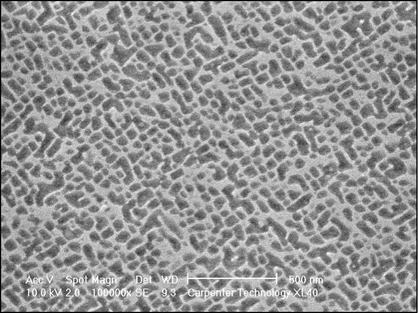
Given the inherent challenges of the extraction approach, Carpenter Technology’s Research and Development (R&D) team has pursued the development of a robust procedure centered around the microscopy method, which relies on capturing in-situ images of the γ’ precipitates. Due to the small size of the particles, electron microscopy in the form of either scanning electron microscopy (SEM) or transmission electron microscopy (TEM) must be used to obtain the necessary resolution to view the particles. Our R&D experts are equipped with a high-resolution field emission-type scanning electron microscope that has the imaging capability necessary to resolve the sub-micron γ’ particles.
Specimen preparation is a key component of the microscopy approach — even the highest performing microscopes cannot compensate for poor preparation. The fundamental goal of our multi-step preparation procedure is to minimize residual deformation that can be easily created during standard metallographic polishing techniques used for light microscopy. A badly prepared surface may appear to be mirror-smooth to the eye, but found to be unusable when imaged in an electron microscope.
Electron Microscope Image Creation and Analysis
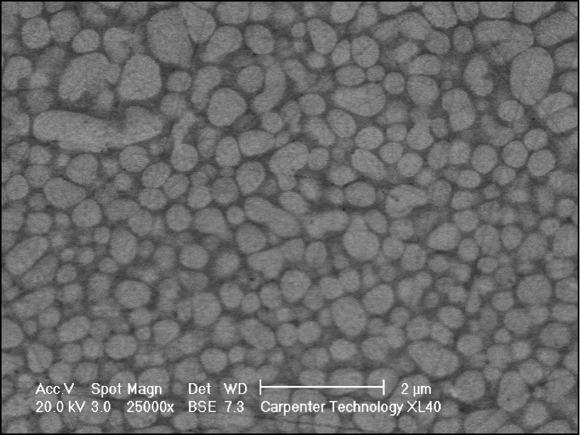
The contrast in an electron microscope image comes from a variety of sources and signals. The two most commonly used signals are secondary electrons and backscattered electrons. Secondary electrons image topographical features on a specimen, while backscattered electrons exhibit a unique characteristic that makes them sensitive to the atomic number of a specimen. This atomic number sensitivity can be used to create images where the contrast reflects changes and variations in the chemical composition within the field of view.
In alloys where the composition of the γ’ precipitates is significantly different than the surrounding metal matrix, the backscattered electron signal can be used to image the particles. In other alloys where the composition of the γ’ phase is not considerably different than the surrounding metal matrix, the specimen surface can be exposed to an agent that acts to selectively attack the γ’ particles, leaving behind pits that reflect the size and shape of the particles that were dissolved. The pits can then be successfully imaged using the topography-sensitive secondary electron signals.
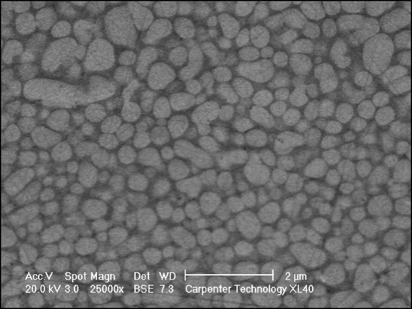
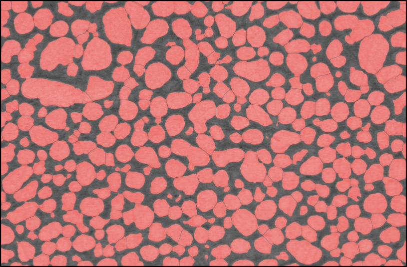
Once a series of high-quality images has been captured, image processing is applied to convert the visual information to quantitative information. Carpenter Technology metallurgists have developed an image analysis algorithm that accurately detects and separates the γ’ precipitates in the image and produces numerical measurements on particle size, shape, and volume fraction.
Real-world applications
This approach has been successfully applied to multiple alloy development projects and has also proven to be a valuable tool in validating the predictions of Carpenter Technology’s mathematical material models. Sharpening and refining these models allows our R&D team to fast-track alloy development by accurately simulating the characteristics of a wide range of material chemistries, thus bypassing the need to perform multiple lengthy and expensive campaigns to physically produce, test, and evaluate series of experimental chemistries.
